Wednesday, October 31, 2018

9th World Congress on Breast Cancer and therapies


Breast cancer is treated both locally (with surgery and radiation) and systemically (hormone therapies, chemotherapy, and targeted therapies).

9th World Congress on Breast Cancer and Therapies (2).png


Local treatment
Local treatment means surgery and/or radiation. You can have a lumpectomy, which removes only the tumour and leaves the healthy part of the breast, or a mastectomy, which removes the whole breast. Your doctor may also remove your lymph nodes, which are under the arm, to make sure there is no cancer there. If you have a mastectomy, you might also decide to have your breast(s) reconstructed. If you have a lumpectomy, you will usually need radiation too.
Systemic treatment
Systemic treatments travel through the bloodstream to attack any cancer cells that spread beyond the breast. Chemotherapy uses drugs to kill cancer cells. Some breast cancer cells depend on hormones like estrogen or progesterone to grow, so women whose breast cancer tumours need estrogen to grow to take hormone (endocrine) therapy.
Targeted therapy attacks only specific types of cancer cells so that normal cells stay healthy; it is used in women who have HER2 positive breast cancer, among others.

Tuesday, October 30, 2018

9th World Congress on Breast Cancer and Therapies


Diagnosing breast cancer is a process. Most women who are diagnosed with breast cancer do not have any symptoms; their cancer is found during routine screening, like a mammogram. If you do have symptoms, or if your mammogram shows something in your breast, tell your doctor as soon as you can. Finding breast cancer when it is still small can give you more treatment options and make you more likely to respond well to them.
Your doctor might also want to remove more tumour tissue and send it for an Oncotype test, a genomic test that gives information about the behaviour of the cancerous tissue. If you've been screened and had abnormal mammogram results, your doctor should do a physical exam, find out your medical history, and tell you if you need further screening to make a diagnosis.
Breast Cancer Summit 2019 (12).png

Diagnostic Mammograms

Sometimes, if you've received abnormal mammogram results, your doctor might need more images to get more information about a suspicious area. S/he may order a diagnostic mammogram, which takes more images than a regular screening mammogram. A radiologist will be present during the diagnostic mammogram to make sure the doctor gets all the images needed. If you originally had a 3D mammogram (also known as 3D tomosynthesis), which takes more pictures than a traditional mammogram, you may not need a diagnostic mammogram and instead have an ultrasound.

Biopsies

When a mammogram shows something suspicious, if no further examination is necessary, a doctor will perform a biopsy. A biopsy removes a few sample cells from the suspicious area on your breast. A kind of doctor called a pathologist looks at the biopsied cells under a microscope and decides if the cells are cancer or not. 80% of the time, the cells are not cancer—they are “benign.”
Finding breast cancer when it is still small can give you more treatment options and make you more likely to respond well to them. If the cells are “malignant,” or cancerous, your doctor might send the tissue sample out for more tests to learn what kinds of treatment your tumour cells might respond to.

Biomarker Tests

Nowadays, the doctors will check biomarkers, molecules that indicate normal or abnormal process taking place in your body, in the tissue taken out as the biopsy. This gives a good picture upfront about the behaviour of a tumour and will help determine the best treatment. Oncotype™ and Mammaprint™ are examples of tests on tumour tissue that can assist in predicting the behaviour of a tumour and the prognosis by analyzing its genetic profile.
Some breast cancer cells depend on hormones like estrogen or progesterone to grow. All cells also have proteins that can be switched on or off; in some breast cancer cells, the HER2 protein is switched on, encouraging the tumour to keep growing. There are treatments specifically for these cells. You may be offered targeted therapy such as anti-estrogen therapy or Herceptin, lapatinib, etc. respectively.

Monday, October 29, 2018

Symptoms of breast cancer may include:

  • Breast lumps
  • Swelling under your arm
  • Pain in your breast or nipple
  • New dimples in the skin on your breast
  • The skin on your breast becoming swollen, red, or hot
  • Sudden changes in your breast size
  • Changes in shape in only one of your breasts
  • Having your nipple newly turn inward (invert)
  • Non-milky liquid coming from your nipple

Not all breast cancer shows itself as a breast lump or through breast pain. In fact, most breast cancer is found in women who did not have symptoms. Still, many women worry that lumps or pain in their breast is definite signs of breast cancer. If you are experiencing unusual breast tenderness, sharp pain in your breast, a burning sensation, painful lumps, or any other symptom, this does not mean you definitely have breast cancer. Diagnosing breast cancer is a process, and the symptoms of a breast tumour can vary from person to person. If you are worried that your pain is a sign of breast cancer, or if you are experiencing any of the above symptoms, talk to your doctor about appropriate next steps

Saturday, October 27, 2018

Breast Cancer Summit 2019

Screening Mammograms 

All women should know the look and feel of their bodies, including their breasts. If, for instance, you have always had an inverted nipple your adult life, it should not be a source of worry. Acute onset, however, is something to bring to your health provider's attention. Knowing the look and feel of your breasts is important in avoiding unnecessary procedures, too. All women should have their doctor do a clinical breast exam on a regular basis.
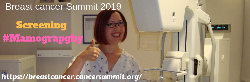
What's a mammogram? Simply, it's still the best way to screen for breast cancer. Mammograms can help find tumours in women who don’t show any symptoms of cancer. But some doctors disagree about screening rules.

Considering having a mammogram? The current guidelines for mammogram screenings are as follows:

  • American Medical Association: annual mammograms at age 40
  • American College of Obstetricians and Gynecologists: annual mammograms at age 40
  • American College of Radiology: annual mammograms at age 40
  • National Cancer Institute: annual mammograms at age 40
  • American Cancer Society: annual mammograms at age 45.
  • American College of Obstetricians and Gynecologists: annual mammograms at age 40.
Most guidelines also say that if a woman doesn’t start having annual mammograms at age 40, annual mammogram screening should start at age 50. Still, there is no one right mammogram screening age.
When you receive your mammogram screening results, talk to your doctor about next steps if they are abnormal. A mammogram does not diagnose breast cancer. Wondering where to get a mammogram? Hospitals, mobile units, and breast centres can all provide mammograms, but you can call ahead to ask about what equipment they use, whether they can provide 3D mammograms or MBI, what insurances they take, and their certification.

3D Mammograms and Molecular Breast Imaging

Breast cancer Summit 2019 8.png
There are special screening rules for people who are at a high risk for breast cancer or who have found a lump or other suspicious area in their breasts. Mammograms can help diagnose the issue, but a doctor may also suggest other tests, like a sonogram or an MRI, because having dense breast tissue makes it harder for mammograms to spot cancer. Dense tissue and tumours both appear light on mammograms, unlike fatty tissue. Women with dense breast tissue may be told to have 3D tomosynthesis (3D mammograms) or molecular breast imaging (MBI), rather than normal mammograms.
3D-tomosynthesis takes multiple X-ray pictures of each breast from many angles and does not compress the breast as much as traditional mammograms. Molecular breast imaging uses a radioactive tracer, injected through a vein, to reveal cancer inside the breast. Breast cancer cells absorb the radioactive substance more than normal cells, so they “light up,” making them easier to spot. A special scanner is used to locate high concentrations of the substance and thereby identify the location of cancer. Unfortunately, molecular breast imaging and 3-D tomosynthesis may not be covered by all health insurance providers. The cost may make them inaccessible to some groups of women, and they are not available in all care facilities. Before you pursue a screening plan, discuss it with your doctor and ask about risks and benefits.

A Note about Mammograms

Some studies and doctors assert that mammograms can lead to overdiagnosis and false positives, which lead women to make difficult choices and engage in expensive and life-altering treatments that could have been avoided or delayed. “Overdiagnosing” means diagnosing cancer that didn’t require immediate treatment without any change to a woman’s prognosis, or diagnosing cancer that never would have required treatment or affected a woman’s health. False positives are suspicious findings that turn out to be normal; they are technically “good news” but can have significant psychological, physical, and economic tolls.

Friday, October 26, 2018

  • Gender - Women are at greater risk than men for developing the disease.
  • Age - Risk increases with age.
  • Genetics - Women with a family history of the disease are at a somewhat greater risk; women with a mutation in specific genes (BRCA1, BRCA2, PALB2, and others that have been identified and can be tested for) are at much greater risk including the risk of developing ovarian cancer. Men who carry the mutation in the BRCA2 gene are at a higher risk for prostate cancer.
  • Having a personal history of breast cancer.
  • Having a prior history of a breast cancer biopsy that revealed a precancerous lesion such as atypia (abnormal cells) or LCIS (lobular carcinoma in situ).
  • A long window of estrogen exposure such as early onset of menstruation and/or late onset of menopause.
  • Previous radiation such as chest radiation for Hodgkins disease.
  • Ethnicity – White women have a higher risk of developing breast cancer than African-American women, but African-American women are more likely to die of the disease at a younger age possibly because of a more advanced stage at diagnosis or having a more aggressive type of the disease (triple negative). Asian, Native-American and Latina women have a lower risk of getting the disease and dying from it.
  • Being overweight or obese (particularly important as you get older).
  • Long term usage of post-menopausal hormone therapy (HRT).
  • Excessive alcohol use, especially more than one drink per day.
  • Lack of physical activity or exercise.
  • Not having children or having children after the age of 35.
  • Not breastfeeding. (Studies suggest long periods of breast feeding may reduce risk of premenopausal or earlier onset breast cancer).
However, it is important to note that having any or all of these risk factors does not mean that you will definitely get breast cancer. 75% percent of the women who develop breast cancer have no identifiable risk factors.
The connection between lifestyle factors and breast cancer, and between environmental toxins and breast cancer, is very important and many researchers are studying this. There is growing evidence that strong links exist. And now there is some evidence that lifestyle modifications may decrease one’s risk of developing a recurrence in those who have had breast cancer.

Thursday, October 25, 2018

9th world congress on Breast Cancer And Therapies

#Breast Cancer #Awareness #October #discount #15% #Join the Summit #Know more about Breast cancer 
Breast cancer is treated both locally (with surgery and radiation) and systemically (hormone therapies, chemotherapy, and targeted therapies).
Local treatment

Local treatment means surgery and/or radiation. You can have a lumpectomy, which removes only the tumour and leaves the healthy part of the breast, or a mastectomy, which removes the whole breast. Your doctor may also remove your lymph nodes, which are under the arm, to make sure there is no cancer there. If you have a mastectomy, you might also decide to have your breast(s) reconstructed. If you have a lumpectomy, you will usually need radiation too.
Systemic treatment
Systemic treatments travel through the bloodstream to attack any cancer cells that spread beyond the breast. Chemotherapy uses drugs to kill cancer cells. Some breast cancer cells depend on hormones like estrogen or progesterone to grow, so women whose breast cancer tumours need estrogen to grow to take hormone (endocrine) therapy.
Targeted therapy attacks only specific types of cancer cells so that normal cells stay healthy; it is used in women who have HER2 positive breast cancer, among others.

Clinical Trials

Chemotherapy (for multiple types of cancer, including triple negative breast cancer)

Hormonal Therapy (for hormone receptor-positive breast cancer)

Radiation

Surgery

Targeted Therapy (for HER2+ breast cancer)

Treatment for Triple Negative Breast Cancer

Final Call for Speakers @breastcancersummit2019

Last Call for participating at the event 9th World Congress on Breast Cancer & Therapies (Breast Cancer Summit 2019) #April 29-30, 2...